Key Points
- A novel molecular engineering technique uses DNA-based microbeads to release growth factors inside organoids, enhancing their complexity and realism.
- The microbeads release their cargo upon UV light exposure, allowing precise timing and location control of signal molecules within the tissue.
- Unlike previous methods, the technique successfully mimicked natural cell compositions when tested on retinal organoids of Japanese rice fish.
- This advancement could accelerate human development, disease, and organoid-based drug discovery research.
A groundbreaking molecular engineering technique has been developed to precisely control the growth of organoids, offering significant advancements in tissue modeling. Researchers from the Cluster of Excellence “3D Matter Made to Order,” along with scientists from Heidelberg University’s Centre for Organismal Studies, the Center for Molecular Biology, the BioQuant Center, and the Max Planck Institute for Medical Research, have introduced DNA-based microbeads that release growth factors or signaling molecules within organoids.
The study was published in the journal Nature Nanotechnology. This innovation leads to the creation of more complex and realistic organoids that closely imitate the natural structure and cell composition of tissues. Organoids are miniature organ-like structures derived from stem cells. They are widely used in basic research to explore human development and disease progression. Previously, controlling the internal growth of these tissue structures was not possible.
Dr. Cassian Afting, a Physician Scientist at the Centre for Organismal Studies, explained, “Using the novel technique, we can now determine precisely when and where in the growing tissue key developmental signals are released.” Tobias Walther, a biotechnologist and doctoral candidate at the Center for Molecular Biology and the Max Planck Institute, emphasized this technique’s precision and control over tissue development.
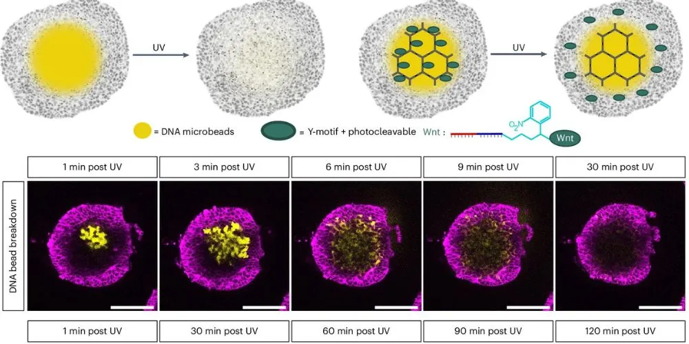
The research team engineered microbeads from DNA, capable of being loaded with proteins or molecules, which release their cargo upon exposure to UV light. This allows researchers to release growth factors at specific times and locations within the organoids. The process was tested on retinal organoids of the Japanese rice fish medaka, where microbeads loaded with a Wnt signal molecule successfully induced the formation of retinal pigment epithelial cells adjacent to neural retinal tissue, closely mimicking natural cell composition.
This method represents a major improvement over traditional approaches, where adding Wnt to culture media induced pigment cells but suppressed neural retina development. The localized release of signaling molecules resulted in a more realistic cell mix, highlighting the potential for creating sophisticated organoid models.
Joachim Wittbrodt, who co-directed the research with Prof. Kerstin Göpfrich, noted, “This opens up new possibilities for engineering organoids with improved cellular complexity and organization,” paving the way for accelerated research in human development, disease, and drug testing.


