

Key Points
- Scientists have developed a new technique to image tiny structures inside human cells more accurately.
- The method combines light microscopy with ion-beam milling and exploits interference of fluorescent light.
- It allows researchers to pinpoint the exact location of a tagged object within a cell.
- The new technique is about ten times more accurate than previous methods.
Getting a clear picture of the tiny machinery inside a human cell is incredibly difficult. One of the best methods, called cryogenic electron tomography (cryoET), involves freezing a cell and then shooting electrons through it to create a 3D image. The problem is, this only works on very thin samples, and most human cells are too thick.
Scientists have a solution for this: they use a beam of ions to shave the cells down into ultra-thin slices. But this creates a new problem: it’s like a game of cellular hide-and-seek. You might have to mill dozens of slices before you find the one that contains the specific structure you’re looking for, like a virus or a particular protein.
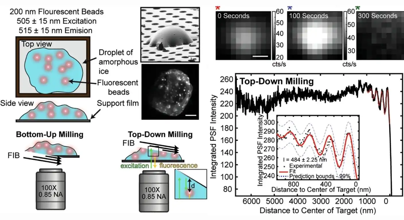
Now, researchers at the SLAC National Accelerator Laboratory have developed a new technique that makes this process much more accurate. By combining light microscopy with ion beam milling, they have found a way to use the “ripples” of fluorescent light to guide the cutting process.
Here’s how it works: they tag the object they want to see with a fluorescent chemical that makes it glow. As the ion beam shaves off the top of the cell, the light from the glowing object shines up. The newly cut surface of the cell reflects that light, causing it to interfere with the incoming light, much like ripples on a pond. This interference causes the fluorescent light to dim and brighten in a predictable pattern.
The researchers wrote software that can read these dimming and brightening patterns and pinpoint the exact location of the tagged object. This allows them to stop the milling process at the perfect moment, ensuring that the structure of interest is perfectly centered in the thin slice.
The new technique is about ten times more accurate than previous methods. To prove it, the team captured a clear image of a tiny, 26-nanometer-wide virus infecting a human cell, a feat previously all but impossible.
Source: Nature Communications (2026).